
Facing eye problems?
Get your eyes examined by our eye specialist team.

WE HELP YOU SEE THE WORLD WITH CLARITY AND COMFORT!
Know Our Story

WE HELP YOU SEE THE WORLD WITH CLARITY AND COMFORT!
Know Our Story

WE HELP YOU SEE THE WORLD WITH CLARITY AND COMFORT!
Know Our Story
WE HELP YOU SEE THE WORLD WITH CLARITY AND COMFORT!
Know Our Story
Our Services

Why choose
Shantilal Shanghvi Eye Institute?

Comprehensive Eye Care Under One Roof
From routine eye check-ups to advanced diagnostic and surgical procedures; high-quality eye care is under one roof.
Modern State-of-the-Art Technology
Equipped with advanced diagnostic and surgical technology for precise diagnoses and treatment that result in improved outcomes and faster recovery times.

Team of Highly Skilled and Experienced Eye Specialists
A well-trained and experienced team of doctors, optometrists, technicians, and nursing staff, that works collaboratively throughout the entire treatment process to ensure that our patients receive the best care.

Empathetic and Equitable Patient Care
Personalized, compassionate care that prioritizes patients' comfort and well-being, irrespective of their background.
Patient Testimonial
AWARENESS IS THE FIRST STEP TOWARDS CURE.
Learn more about eye diseases, available treatments, and potential technological advancements.
Know More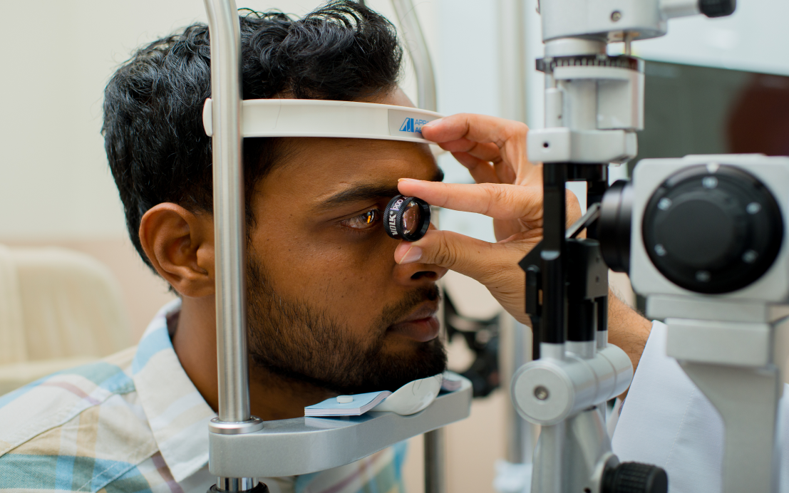
Contact Us
A Unit of Shantilal Shanghvi Foundation
Copyright © JGDHealth. All Rights Reserved.