
Case Stories
Successful Management of Retinal Detachment
Without Surgery.
- Dr. BHAGYASHREE MESHRAM
Other Stories
Bright Beginnings - A 3-month-old child’s
journey to visual restoration
- Dr. JENIL SHETH
A 10-year-old boy from a Mumbai slum was
brought to the Shantilal
Shanghvi Eye Institute.
- DR. NIDHI AGARWAL
A Miracle Journey of a Premature Baby's
Triumph
over Retinopathy of Prematurity
- Dr. SUSHMA
JAYANNA
HealEYE
Health Insights

Dr. BHAGYASHREE MESHRAM
Faculty
Retina and ROP Services
Successful Management of Retinal Detachment Without Surgery.
Case History
A 72-year-old female presented to our OPD with complaints of seeing black spots in her left eye. She had undergone cataract surgery two months prior. On examination, she was diagnosed with left-eye retinal detachment, associated with a small break in the retina. Her vision was 20/20 at the time of presentation. Since retinal detachment can progress toward the macular region, leading to rapid vision loss, immediate intervention was necessary to preserve her sight.
Treatment
The patient underwent pneumatic retinopexy on the same day an OPD-based procedure that involves injecting gas into the eye to help reattach the retina. Strict positioning compliance was essential for the success of this procedure. During follow-ups, laser photocoagulation was performed around the retinal break to reinforce its stability and prevent further detachment.
Result
With excellent compliance in maintaining the advised position and attending multiple follow ups,the patients retina remined stable and surgical intervention was successfully avoided. This case highlights the significance of early diagnosis, timely intervention, and patient adherence in effectively managing retinal detachment.
Pneumatic Retinopexy (PnR) is a minimally invasive, office-based procedure used to treat certain types of retinal detachments. It involves injecting a gas bubble into the vitreous cavity of the eye, which helps push the detached retina back into place. The patient must maintain a specific head position to ensure the bubble effectively position to ensure the bubble effectively seals the retinal break. Once reattached, laser photocoagulation or cryotherapy is applied to secure the retina and prevent further detachment. PnR is a quick and effective alternative to surgery, offering faster recovery and reduced risks when performed on suitable cases.
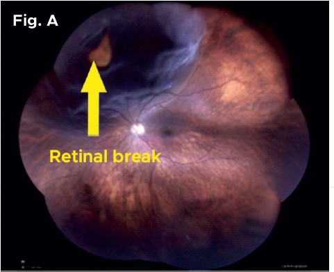
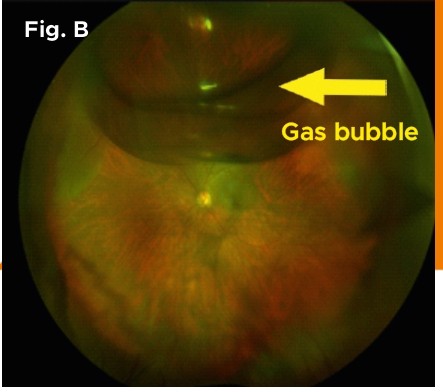
The fundus images illustrate the Outcome:
- Fig. A: The top left part of the retina (the light-sensitive layer inside the eye) has a tear, causing it to lift and fluid to collect underneath.
- Fig. B: On the first day after a gas bubble was placed in the eye, the retina is almost back in its normal position.
- Fig. C: The retina is fully reattached, with laser spots placed around the tear to keep it secure.
- Fig. D: A joyful patient with her daughter-in-law!

Contact Us
A Unit of Shantilal Shanghvi Foundation
Copyright © JGDHealth. All Rights Reserved.